Management of Hypodontia with canine substitution: A Case Report
- Dr. Tariq Ansari, Assistant Professor Department of Orthodontics and Dentofacial Orthopedics, Yenepoya Dental College, Yenepoya University, Mangalore
[email protected]
- Dr.Rohan Mascarenhas. Professor, Department of Orthodontics and Dentofacial Orthopedics, Yenepoya Dental College, Yenepoya University, Mangalore
- Dr. Husain C., M.D.S. Former Post graduate student , Department of Orthodontics and Dentofacial Orthopedics, Yenepoya Dental College,Yenepoya University, Mangalore
- Dr. Akhter Husain, M.D.S. Professor and Head, Department of Orthodontics and
Dentofacial Orthopedics, Yenepoya Dental College, Yenepoya University,
ABSTRACT :
Introduction: Management of hypodontia especially involving upper lateral incisors in orthodontic patients can pose a challenge in orthodontic management. Canine substitution is a clinically accepted method wherein the canines are moved into the position of the lateral incisors and subsequently their morphology is altered to resemble the lateral incisor .Methodology: An 18 year old female patient exhibited missing maxillary lateral incisors bilaterally, midline diastema and spacing in the upper anterior teeth. Pre-treatment radiographs (OPG and Cephalograms) were taken. The treatment plan involved substitution of the missing lateral incisors with the maxillary canines. The patient was treated with the MBT technique. Finally post treatment records were made and compared to the pre-treatment records. Results: The canines have been moved to the lateral incisor position and they have been converted to resemble lateral incisors with satisfactory results. All dentoalveolar and skeletal changes have been brought about without any additional intra oral or extra oral device. Conclusion: Correction of anterior spacing along with proclination and missing lateral incisors can be achieved with canine substitution with satisfactory aesthetics.
Keywords: Hypodontia , Canine substitution, Midline Diastema ,
TRATAMIENTO DE LA HIPODONCIA CON SUSTITUCIÓN CANINA, CASO CLÍNICO
RESUMEN:
Introducción: el tratamiento de hIpodoncia involucra a incisivos laterales superiores en pacientes de ortodoncia, pudiendo plantear un desafío en el manejo ortodóncico de estos pacientes. La substitución canina es un método clínicamente aceptado en donde los caninos son movidos en la posición de las incisivos laterales y posteriormente su morfología es cambiada para parecerse a la incisivo lateral. Método: un paciente femenino de 18 años presentó ausencia bilateral de incisivos laterales maxilares, diastema en línea media y espaciamiento de los dientes anteriores superiores. Las radiografías de pretratamiento (Panorámica y Cefálica) fueron tomadas. El plan de tratamiento implicó la substitución de los incisivos laterales ausentes con los caninos maxilares. El paciente fue tratado con la técnica MBT. Finalmente se tomaron los registros al final del tratamiento y estos se compararon con los de pretratamiento. Resultados: los caninos han sido movidos a la posición de incisivo lateral y han sido cosméticamente tratados para parecerse a incisivos laterales con resultados satisfactorios. Todos clos cambios dentoalveolares y esqueléticos han sido realizados sin ningun otro dispositivo extraoral o intraoral suplementario adicional. Conclusión: la Corrección del espaciado anterior junto con proclinacion y ausencia de incisivos laterales puede ser conseguida mediante la substitución canina con la preparación estética satisfactoria.
Palabras clave: Hipodoncia, substitución Canina, Línea media Diastema |
The incidence of missing teeth in the oral cavity has been considered as a progressive step in evolution (1). According to a theory proposed by Butler known as the Butler Field Hypothesis the most mesial tooth in an arch is the most morphologically stable tooth and the abnormal morphological variation increases progressively to the most distal tooth in the arch (2). This theory was proposed for mammals and was adapted by Dahlberg who applied this principle for human beings and divided the human dental arch into different developmental fields and said that the mesial tooth in each developmental field is the most morphologically stable tooth in the arch(3).
Management of hypodontia especially involving upper lateral incisors in orthodontic patients can pose a challenge in orthodontic management. Management of cases with missing maxillary lateral incisors has many options which includes the replacement of missing teeth with the help of a prosthesis or implant both of which have their drawbacks According to. Dr. Kokich the most aesthetic and functional treatment option for management of a missing maxillary lateral incisor is canine substitution . Though the canine can be re shaped to have an aesthetic appearance of a maxillary lateral incisor, sometimes a Crown or a veneer may have to be placed on the substituted canine to improve easthetics (4).Correction of proclination of the anteriors along with canine substitution would require the protraction of the maxillary posterior segment in cases which exhibit class I molar relationship.
Methodology:
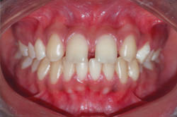
An 18 year old female patient exhibiting a class I molar relation on the left side and an End-on molar relationship on the right side as well as an End on canine relationship bilaterally reported to the department of Orthodontics at Yenepoya University. This patient also exhibited missing maxillary lateral incisors bilaterally, midline diastema and spacing in the upper anterior teeth and rotated maxillary premolars.The patient had satisfactory periodontal health, good bone support and adequate attached gingiva, she also had good oral hygiene.
Routine records of the patient were made such as
Detailed case history
Pre-treatment study models
Extra oral and intra oral photographs
Lateral cephalograms
Orthopantomograms
Intra-oral periapical radiographs were acquired.
On radiographic investigation it was determined that the patient exhibited hypodontia with respect to the maxillary lateral incisors.
 |
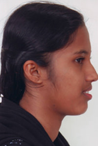 |
Fig
1
Pre treatment extra oral frontal smile |
Fig.2
Pre treatment extra oral profile |
 |
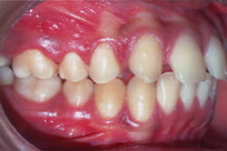 |
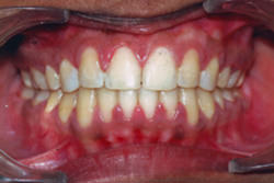
Fig.3
Pre-treatment Intra-oral frontal view |
Fig.4
Pre-treatment Intra-oral right side view |
 |
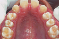 |
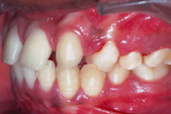
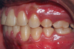
Fig.5
Pre-treatment Intra-oral left side view |
Fig.6
Pretreatment Maxillary arch occlusal view |
 |
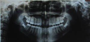 |
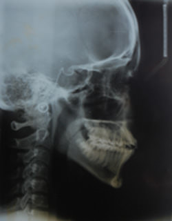
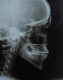
Fig.7
Pre treatment lateral cephalogram |
Fig.8
Pre treatment O.P.G |
The treatment plan involved substitution of the missing lateral incisors with the maxillary canines . The patient was treated with the MBT technique (.022 inch slot). The leveling and aligning was carried out with 0.016" HANT (Heat Activated Nickel Titanium) and 0.019" x 0.025" HANT arch wires.
Stainless steel 0.019" x 0.025" rectangular wires were used for complete tip and torque expression and also for space closure as recommended by the MBT technique
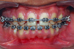
Initially only the maxillary arch was bonded.(Fig 9) Lingual buttons were bonded to the palatal surfaces of the maxillary first premolars, lingual buttons were also welded to the palatal surfaces of the maxillary molars . Then an elastic thread was tied between the lingual buttons of the maxillary first premolars and also the lingual buttons of the maxillary first molars to bring about derotation of the rotated maxillary first premolars.(10)
 |
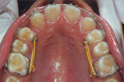 |
Fig.9
Appliance bonded to the maxillary arch. |
Fig.10
Derotation of Premolars |
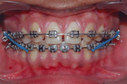
After the premolars were de rotated the 016 round arch wire was replaced with 019x025 NiTi wires followed by the 019x025 stainless steel wire arch wire . Simultaneously the Mandibular arch was also bonded.(Fig.11) An elastic chain was placed extending from the maxillary first premolar of one side to the maxillary first premolar on the opposite side in order to close the anterior spacing and the midline diastema. This brought about a mesialisation of the maxillary canines into the place of the missing lateral incisors(12)
 |
 |
Fig
11
Appliance bonded to Mandibular arch |
Fig.12
Mesialisation of maxillary canines |
Once space developed distal to the maxillary first premolar class III
elastics were placed between the maxillary first molars and the distal hook of
the Mandibular cuspid hooks(Fig.13)
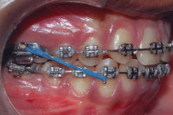 |
Fig.13
Intermaxillary Class III elastics placed bilaterally |
This brought about a mesialisation of the posterior segment along with the maxillary first molar to a class II molar relationship bilaterally .The maxillary canines have been altered to conform to the shape of the lateral incisors and the maxillary first premolars have been altered to resemble the maxillary canine.
The cephalograms are evaluated for inclination and position of the upper incisor and lower incisors.
Results:
The post treatment evaluation shows an acceptable overjet and overbite. The bucal segment shows a class I canine relationship bilaterally and a class II molar relationship bilaterally.
 |
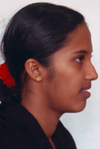 |
Fig.14
Post treatment extra oral frontal smile |
Fig.15
Post treatment extra oral profile |
 |
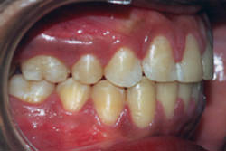 |
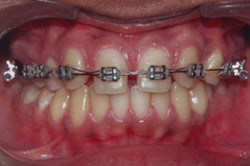
Fig.16
Post-treatment Intra-oral frontal |
Fig.17
Post-treatment Intra-oral right side |
 |
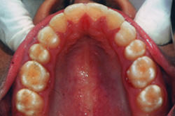 |
Fig.18
Post-treatment Intra-oral left side |
Fig.19
Prost treatment Maxillary arch occlusal |
 |
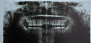 |
Fig.20
Post treatment lateral cephalogram |
Fig.21
Post treatment O.P.G |
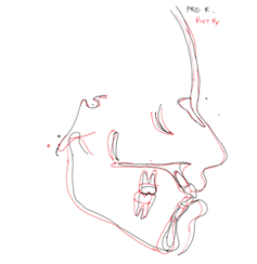 |
Fig.22
Pre-treatment and post-treatment lateral cephalogram superimposition.
Black denotes pretreatment tracing. Red denotes post treatment |
Conclusion:
Proclination of teeth, a lack of space and a class III skeletal tendency accompanied with missing maxillary lateral incisors needs an individualised treatment plan. Protraction of the maxillary buccal segment is one of the methods for gaining space and has been used here effectively to bring about desired changes. The canines have been moved to the lateral incisor position and they have been converted to resemble lateral incisors with satisfactory results. All dentoalveolar and skeletal changes have been brought about without any additional intraoral or extra oral device.
References
- Silverman NE , Ackerman JL 1979 Oligodontia : A study of its prevalence and variations in 4032 children . Journal of Dentistry for children 46:470-477
- Butler PM 1939 Studies of the mammalian dentition . Differentiation of the post- Canine dentition . Proceedings of the Zoological Society of London .109:1-36.
- (Dahlberg AA 1945 The changing dentition of man . Journal of the American Dental Association.
- Kokich, V., & Kinzer, G. (2005). Managing congenitally missing lateral incisors. Part I: Canine substitution., 17(1), 5-10
|
