Introduction
Class III malocclusion is a subject of interest and concern for orthodontists both in research and clinical practice. While the dental class III malocclusion may not have any significant skeletal discrepancy, the skeletal class III malocclusion is associated with a wide variety of underlying skeletal and dental patterns. (Baratali &al., 2007)(1)
The prevalence of skeletal class III malocclusion varies among different races and populations. The highest prevalence is among Asians of the Far East and the lowest is in Caucasians.(Franchi & al.,2005)(2)
Craniofacial characteristics of growing subjects with Class III relationships have been mostly studied in the sagittal and vertical plane. But the skeletal malocclusions are complex clinical entities that comprise of three-dimensional dental and skeletal components. (Franchi & al., 2005)(2)
The transverse component of sagittal skeletal disharmonies presents clinical features that point to the need for maxillary expansion prior to correction of the anteroposterior discrepancy in growing subjects. (2) Bilateral facial asymmetries and development of the oronasal area can be better assessed from a transverse analysis of Postero-Anterior (P-A) cephalometric radiographs. (Dawn Wagner & al., 2005)(3)
Unfortunately, studies of the transverse relationship of the maxilla to the mandible in Class III subjects in the mixed dentition have been limited to the analysis of the arch widths which are measured on dental casts. There is no information available for transverse dimensions in Class III subjects. (Franchi & al., 2005)(2)
Class III skeletal malocclusion though primarily a sagittal jaw discrepancy, it would be interesting to study the skeletal features in transverse plane also. It is therefore essential to evaluate the skeletal relationship in all three planes of space. Hence this study was designed and conducted with the objective of assessment of facial asymmetry in transverse plane in individuals having skeletal class III jaw discrepancy.
Materials and methods
90 subjects (45 males and 45 females) between 18 and 30 years of age were selected as per the following criteria.
Inclusion criteria for study group
- All intact permanent dentition (excluding 3rd molar)
- Clinically obvious maxillary deficiency/ mandibular excess
- Individuals with Class III profile
Exclusion criteria
- Skeletal abnormalities like cleft lip and palate and other cranio facial deformities
- Prior orthodontic /surgical treatment
- Deviation of mandible on opening and closing
Inclusion criteria for the control group
The subjects for the control group were selected based on their pleasing class I profile, normal dental occlusion with normal overjet and overbite with no midline deviations.
Written informed consent was obtained from each individuals and the project was approved by the Institutional Review Committee. Lateral cephalograms were made and the subjects were classified based on their sagittal relationship as follows.
Group I: Control group: 30 Individuals with class I malocclusion (15-Males, 15-Females) with SNA=82+-2 and SNB=80 +-2
Group II: 30 individuals (15-Males, 15-Females) with Skeletal class III with maxillary deficiency SNA<84, N PER A (II HP) <-6mm, SNB=80+-2
Group III: 30 individuals (15-Males, 15-Females) with Skeletal class III with mandibular excess SNA= 82+-2, N PER B (II HP)>4mm and SNB> 82
Postero-Anterior (P-A) cephalograms were made for all the selected subjects under standardized conditions and were traced on 0.03 acetate paper using 2H pencil by a single operator.
The landmarks were identified for analysis (Grummons &al., 1987)4 in Postero-Anterior cephalometric tracing (Fig 1) and skeletal asymmetry analysis was carried out (Fig 2)
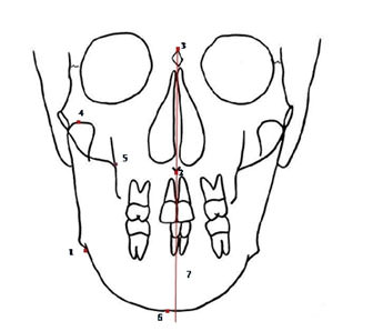
Fig.1
Landmarks identified
- Antegonial Notch (Ag) - Highest point of the notch of the lower border of mandible
- Anterior nasal spine (ANS)- The most anterior point on the maxilla at the level of the palate.
- Crista Galli (Cg) - Neck of Crista galli.
- Condylion (Co) -The most posterior superior point on the condyle of the mandible.
- Jugal process (J) -Medial aspects of the jugal process.
- Menton (Me)- The most inferior point on the symphysis of the mandible, as seen on the lateral jaw projection.
- Gonion (Go)- The point on the contour of the mandible determined by bisecting the angle formed by the mandibular and ramal planes.
- Mid Sagittal Reference Line (MSR) -Vertically fom Cg through ANS to the chin area.
Measurements used in the study (Fig 2) are as follows
- Mandibular Morphology
Left - right triangles are formed from the heads of the condylar processes or condylion (Co), Antegonial notch (Ag) and Menton (Me). These are split by ANS-Me line and compared.
- Volumetric Comparison
Two volumes are calculated from the area defined by each Co-Ag-Me and the intersection with a perpendicular from Co-MSR.
- Maxillo - Mandibular Comparison of Asymmetry
Perpendiculars are drawn to MSR from J and Ag and connecting lines from Cg-to J and Ag. This produces 2 pairs of triangles, each is bisected by MSR.
- Linear Asymmetries
The linear distance is measured from MSR to Co, J, Ag and Me.
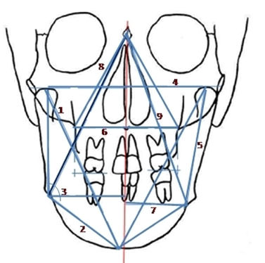
Fig.2
Measurements carried out
Statistical analysis
The mean and standard deviation for each measurement was calculated. Paired t-test was used to test the significance (p= 0.01 or less) in the difference between the right and left sides of the face and for any gender difference.
Results
The facial symmetry of skeletal class III individuals was analyzed using Grummon's analysis in a frontal cephalogram and the following results were obtained.
Mandibular Morphology (Table 1, 2, 3)
No significant differences were observed between right side and left side values in relation to Co-Ag, Ag-Me and gonial angle in both in males and females of all the three groups
Volumetric Comparison (Table 1, 2, 3)
Table 1
Mandibular morphology and volumetric comparison for group I
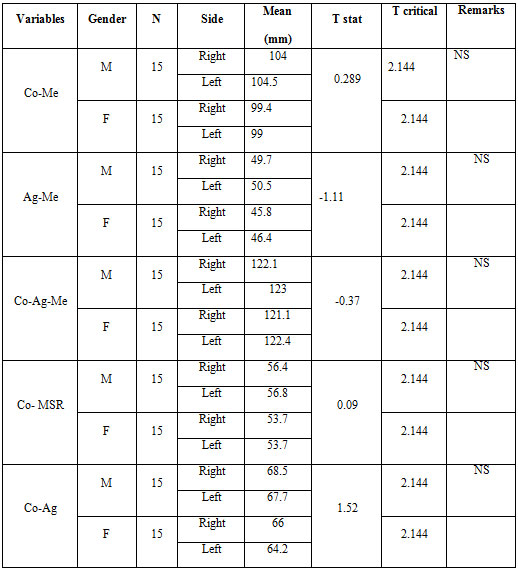
Table 2
Mandibular morphology and volumetric comparison for group II
(Maxillary Deficiency)
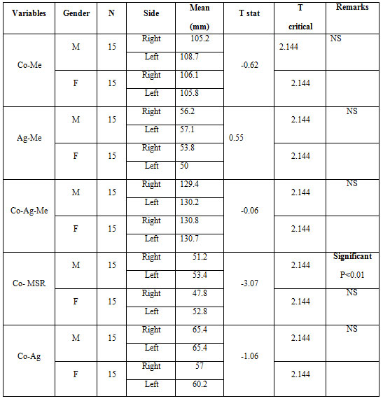
Table 3
Mandibular morphology and volumetric comparison for group III
(Mandibular Excess)
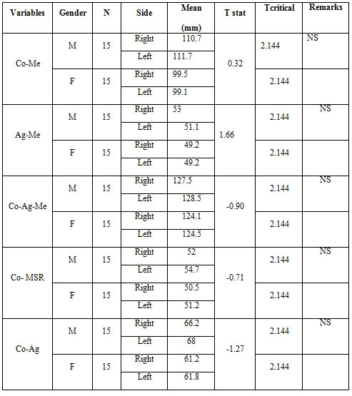
No significant differences were observed between right side and left side values in relation to Co-MSR, Co-Ag and Ag-Me in both males and females of all three groups
Maxillo-Mandibular Comparison of Asymmetry (Table 4, 5, 6)
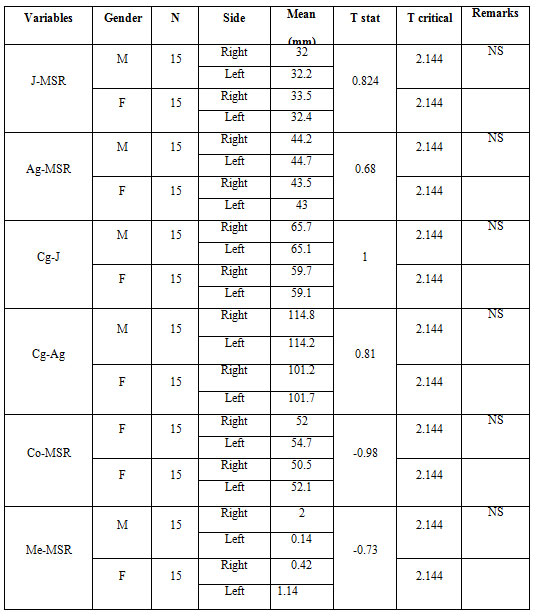
Statistically significant differences were observed between right and left side values in relation to Ag-MSR in females of group II individuals. No significant differences were observed between right side and left side values in relation to Cg-J, Cg-Ag, J-MSR Group I Group II and Group III.
Linear Asymmetries (Table 4, 5, 6)
Statistically significant difference was observed between right and left side values in relation to MSR-Co in males and MSR-Ag in females of group II individuals. In males, no significant differences were observed between right side and left side values in relation to MSR -J , MSR- Me in group I , group II and group III,
Table 4
Maxillo-mandibular comparison and linear measurements for group
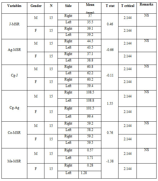
Table 5
Maxillo-mandibular comparison and linear measurements for group II (Maxillary Deficiency)
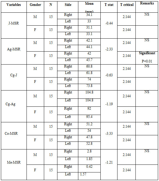
Table 6
Maxillo-mandibular comparison and linear measurements for group III (MandibularExcess)
Discussion
Craniofacial characteristics of growing subjects with either Class II or Class III skeletal relationships have been mostly studied in the sagittal and vertical plane. But both malocclusions are complex clinical entities that comprise of three-dimensional dental and skeletal components. (Baratali &al., 2007)(1)
Unfortunately, studies of the transverse relationship of the maxilla to the mandible in Class III subjects have been limited to the analysis of the arch widths which are measured on dental casts. There is no information available for facial transverse dimensions in Class III subjects. (Baratali &al., 2007)(1)
Most of the normative data have been based on sagittal aspects of dentoskeletal structures with the current emphasis on orthodontic diagnosis obtained from information from the postero-anterior cephalometric radiograph films. However, evaluation also is needed in the transverse dimension for a comprehensive dentofacial evaluation. (DawnMW & al, 2005)(3)
Hence this study was planned and designed for the assessment of skeletal symmetry in skeletal class III individuals. The data obtained would give us an insight into the skeletal relationships in the transverse plane in these individuals.
Postero-anterior cephalograms were used to assess skeletal asymmetry. PA view is a valuable tool in the study of right and left structures since they are located at relatively equal distance from the film and X-ray source, as a result the effect of unequal enlargement by the diverging rays is minimized and the distortion is reduced. Comparison between sides is therefore more accurate since the midlines of the face and dentition can be recorded and evaluated. (Bishara SE & al, 1994)(5)
The lower usage of PA cephalograms may be attributed to the fact that orthodontic educational centers do not emphasize the importance of PA cephalometric evaluation or the difficulties encountered in conducting such evaluation. These problems include the errors associated with reproducing head posture and identifying landmarks of the structures that are superimposed or not identifiable with poor radiographic technique, as well as concern about additional exposure to radiation.(CassidyKM & al,1998)(6)
There are many types of postero-anterior analysis used for assessment of the facial asymmetry like Svanholt and Solow analysis (Svanholt and Solow, 1977), Grayson analysis (Grayaon & al, 1983), Hewitt analysis (Hewitt, 1975) and Ricketts analysis. For the present study Grummons analysis was used for the assessment of the asymmetry. Analysis proposed by Grummons and Kappeyne Van De Cappello (1987)4 contains quantitative assessment of vertical dimensions and proportions. This is a comparative and quantitative postero-anterior analysis. This type of analysis provides a practical, functional method of determining the location and amount of facial asymmetry. (Peck & al, 1991)(7)
In the present study the following components of Grummon's analysis were used - Mandibular Morphology, Volumetric Comparison, Maxillo-Mandibular Comparison of asymmetry, Linear asymmetry assessment and Maxillo-Mandibular relation.
Individuals in the age group of 18 years to30 years were selected as per the inclusion criteria and exclusion criteria. Lateral cephalogram, frontal cephalogram and were taken after obtaining the written consent. 30 individuals (15 males and 15 females) with maxillary deficiency and 30 individuals (15 males and 15 females) with mandibular were considered for the study.
10 cephalometric measurements were made to determine and evaluate the dentoskeletal characteristics in transverse plane in skeletal class III malocclusion.
The mean and standard deviation for each measurement was calculated. Paired t-test was used to test the significance in the difference between the right and left sides of the face and for any gender difference.
Statistically significant differences were observed in relation to Ag-MSR in females having maxillary deficiency. No significant difference was observed between right side and left side values in relation to Cg-J, Cg-Ag, J-MSR in all three groups of individuals. This finding is in agreement with studies by Rossi M & al,(2003) (8) Haraguchi & al,(2002) (9) and Server TR and Profit, (1997)(10) but is in contradiction to studies by Shah and Joshi,(1991)(11) according to which there is a tendency for the maxilla to be more asymmetric than mandible.
Statistically significant differences were observed in relation to MSR-Co in males and MSR-Ag in females having maxillary deficiency. No significant differences were observed between right side and left side values in relation to MSR -J, MSR-Me in all groups of individuals.
The findings of this study show a tendency for the mandible to be more asymmetric. This may be because (1) the mandible grows longer than the maxilla and thus is likely to show more deviation and (2) the mandible is a mobile apparatus whereas the maxilla is connected rigidly to its adjacent skeletal structures.(ServerTR &al,1997)(10)
In the present study, consistent left side dominance has been found in all cephalometric measurements both in males and females. . This finding is similar to a study done by Giovanoli & al,(2003)(12) who had reported left sided dominance. However, Haraguchi& al,(2002)(9) Shah and Joshi,(1978)(11) Peck & al,(1991)(7) in their asymmetry analysis, reported a right side dominance.
The present study revealed significant skeletal asymmetry in transverse plane in individuals with skeletal class III malocclusion. This fact must be taken into account during diagnosis and treatment planning.
Further studies with larger sample size comprising of different skeletal and dental malocclusions in various racial groups at different age groups will be required for assessment of skeletal and dental asymmetries.
Conclusion
The following conclusions can be drawn from the study
- Variations in facial symmetry were evident between the right and left sides in individuals with skeletal class III malocclusion.
- The mandible is found to be more asymmetric than maxilla in patients with maxillary deficiency.
- Volumetric comparison showed male dominance in individuals with maxillary deficiency and maxilla-mandibular relation showed female dominance in maxillary deficiency group.
Reference
- Baratali Ramezanzadeh,Maryam Pousti,Mansour Bagheri(2007).Cephalometric evaluation of dentofacial features of class III malocclusion in adults of Mashad ,Iran. .JODD;3:125-30
- Franchi L,Baccetti T (2005). Transverse maxillary deficiency in class II and III malocclusions :a cephalometric and morphometric study on postero-anterior films . Orthodontic craniofacial research. 8:21-28
- Dawn M.Wagner and Chun-Hsi Chung (2005). Transverse growth of the maxilla and mandible in untreated girls with low, average, and high MP-SN angles: A Longitudinal study. Am J Orthod Dentofacial Orthop; 128:716-723.
- Grummons DC, Kappeyne (1987). A frontal asymmetry analysis. J Clinical Orthod; 21: 448 -65
- Samir E.Bishara, Pauls.Burkey, JohnG.Kharouf(1994). Dental and facial asymmetries: A review. Angle Orthod; 64(2):89-98.
- Kelvin M Cassidy, Edward F Harris, Elizabeth A. Tolley, Robert Keim (1998). Genetic influence on dental archform in orthodontic patients. Angle Orthod ;68(50):445-454
- Peck, Leena Peck, Matti Kataja (1991). Skeletal asymmetry in esthetically pleasing faces Sheldon. Angle Orthod; No. 1: 43 - 48.
- RossiM, Ribeiro E, Smith R ( 2003). Craniofacial asymmetry in development: An Anatomical study. Angle orthod ; 73:381
- Seiji Haraguchi. Kenji Takada, Yoshitaka Yasuda (2002). Facial asymmetry in subjects with skeletal class III deformity. Angle Orthod; 72: 28-35
- Server TR, Proffit WR (1997). The prevalence of facial asymmetry in the dentoalveolar deformities population at the University of North Carolina. Int J Orthod Orhognath Surg ; 171:12
- Shah SM. Joshi MR (1978). An assessment of asymmetry in the normal craniofacial complex. Angle Orthod; 48(2):141-48.
- Giovanoli.p, Tzou CHU, PionerM (2003). Three dimensional video analysis of facial movements in health volunteers, Br.J plast Surg, 56:644.