

INTRODUCTION
Dental impaction is the retention of a tooth within the maxillary or mandibular bone beyond the expected eruption time, usually due to physical barriers such as adjacent teeth, bone, or soft tissue.[1] Permanent canines are most frequently affected, especially in the maxilla, due to their complex eruptive path.[2,3] Prevalence ranges from 1% to 5%, with female predominance.[1,4] Impaction may cause root resorption, malalignment, cysts, and functional/esthetic impairment, supporting early diagnosis.[2] Etiology is multifactorial, including local, systemic, and genetic factors.[5,6] Familial clustering suggests heritability.[7,8] Management often requires interdisciplinary care and surgical–orthodontic intervention.[9] This study analyzes prevalence and radiographic patterns in northeastern Brazil.
MATERIALS AND METHODS
This was a retrospective, descriptive, study conducted through the analysis of panoramic radiographs from patients treated at a public university dental clinic in northeastern Brazil between 2017 and 2025. The study was approved by a local Research Ethics Committee (CAAE: 28805020.7.0000.0055), approved on 15 March 2021.
Population and Sample
A total of 4,954 panoramic radiographs obtained during the study period were evaluated. After applying the inclusion and exclusion criteria, 4,053 radiographs were deemed eligible. Of these, 52 radiographs presenting at least one impacted canine were selected, constituting the final sample.
Inclusion and Exclusion Criteria
Patients aged between 10 and 65 years who presented one or more impacted canines identified on panoramic radiographs were included. Patients with a history of orthodontic treatment and those who had undergone previous canine extraction were excluded.
Procedures and Data Collection
Radiographs were acquired using a digital panoramic X-ray unit (EAGLE, Dabi Atlante), operating at 85 kVp and 10 mA. Image analysis was performed independently by two previously calibrated examiners, with an interexaminer agreement index greater than 90%. Calibration was carried out through the evaluation of 20 radiographs not included in the sample, ensuring standardization in the interpretation of findings.
The variables analyzed included:
Location of impacted canines (maxilla or mandible);
Laterality (right or left);
Position of impacted maxillary canines according to the classification proposed by Yamamoto et al.(11);
Transmigration pattern of impacted mandibular canines according to the classification proposed by Mupparapu(12);
Depth level of impacted mandibular canines according to the classification proposed by Yavuz et al.(13);
Presence of associated dental and radiographic alterations.
Classification of Impacted Canines
In this study, dental impaction was defined as the retention of a tooth that is prevented from erupting into its normal position due to physical obstacles, such as other teeth, bone tissue, or soft tissue present along its eruptive path.(10)
Yamamoto et al. (11) proposed a classification system for canine impaction based on tooth location as observed on panoramic radiographs [Figure 1].
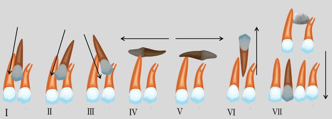
Figure 1. Yamamoto classification of maxillary impacted canines.
Mupparapu (12) proposed a classification system to categorize intraosseous transmigration and ectopic eruption of mandibular canines. This classification is based on the migration pattern of the tooth and its position in the mandible relative to the dental midline, establishing five distinct types:
Type I: Canine positioned mesioangularly near the midline, labial or lingual to the anterior teeth.
Type II: Canine impacted horizontally near the inferior border of the mandible, inferior to the apices of the incisor teeth.
Type III: Canine erupting on the contralateral side.
Type IV: Canine impacted horizontally near the inferior border of the mandible, below the apices of the posterior teeth on the contralateral side.
Type V: Canine positioned vertically at the midline, with the long axis of the tooth crossing the midline.
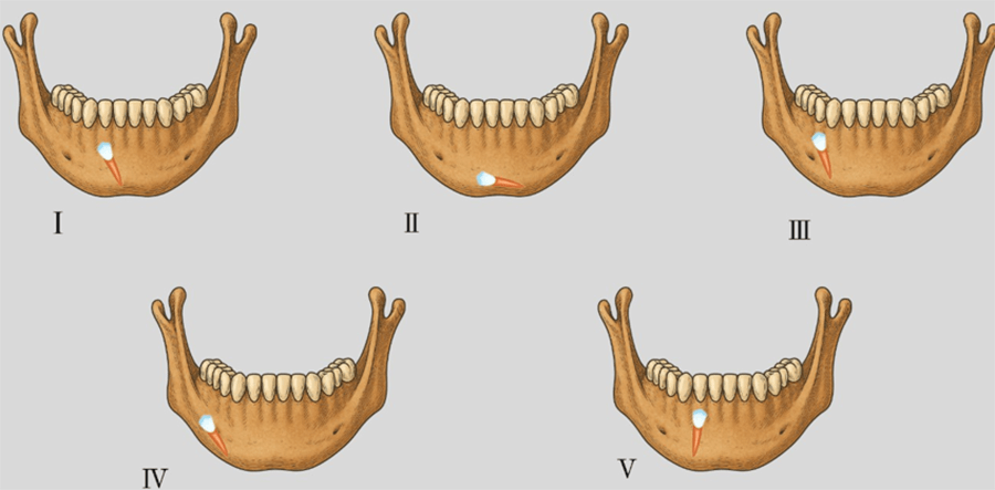
Figure 2. Mupparapu classification of transmigrated mandibular canines.
For mandibular canines, Yavuz et al. (13) proposed a classification based on the depth of dental impaction. According to the authors:
Level A: The crown of the impacted tooth is positioned at the level of the cementoenamel junction of adjacent teeth.
Level B: The crown is located between the cervical line and the root apices of adjacent teeth.
Level C: The crown of the impacted canine is located below the root apices of adjacent teeth.
Statistical Analysis
Data were tabulated using Microsoft Excel software and analyzed descriptively. Categorical variables were expressed as absolute and relative frequencies. The prevalence of canine impaction was calculated as the number of patients with at least one impacted canine divided by the total number of eligible patients, multiplied by 100.
The results were presented in tables and charts according to sex, location, laterality, depth, and the radiographic classifications adopted.
Use of Artificial Intelligence
ChatGPT (OpenAI) was used exclusively to support the linguistic and grammatical revision of the manuscript, including improvements in clarity, readability, and academic English. The tool was not used for data collection, data analysis, interpretation of results, or generation of scientific conclusions. All AI-assisted revisions were critically reviewed and approved by the authors, who take full responsibility for the accuracy and integrity of the final content.
RESULTS
A total of 4,954 panoramic radiographs obtained between 2017 and 2025 were analyzed. After application of the inclusion and exclusion criteria, 4,053 radiographs were considered valid, comprising patients aged 10 to 65 years, of whom 2,324 (57.34%) were female and 1,729 (42.66%) were male.
The prevalence of canine impaction was 1.28%, corresponding to 52 patients who presented at least one impacted canine, totaling 81 affected teeth. The prevalence was slightly higher in females (1.33%) compared with males (1.27%).
Impacted canines were predominantly located in the maxilla, accounting for 93.83% of the teeth (n = 76), whereas 6.17% (n = 5) were located in the mandible. Regarding laterality, a higher occurrence was observed on the right side (54.43%) compared with the left side (45.57%) (Table 1).
Table 1. Percentage distribution of impacted canines according to dental arch and side. Jequié, Bahia, Brazil, 2025.

Table 2. Classification of impacted canines in the maxillary arch according to Yamamoto et al. (2003). Jequié, Bahia, Brazil, 2025.
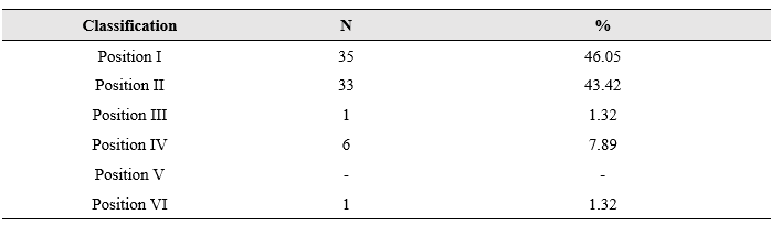
The vertical position (Position I) was the most prevalent among maxillary impacted canines, accounting for 46.05% of cases, followed by the mesioangular position (Position II), with 43.42%. The lowest frequencies were observed in Position VII (ectopic/labiopalatal) and Position III (distoangular), both representing 1.32% of cases. No cases were recorded in Position V (horizontal).
All cases of mandibular transmigration were classified as Type I (100%) according to the classification proposed by Mupparapu (12), which is characterized by a mesioangular position of the canine, with the crown crossing the midline and being located on the labial or lingual aspect of the anterior teeth.
Table 3 presents the classification of the depth level of impacted mandibular canines according to the classification proposed by Yavuz et al..(13)
Table 3. Depth level of impacted mandibular canines according to Yavuz et al. (2007). Jequié, Bahia, Brazil, 2025.

Among the impacted mandibular canines, most were classified as Level B (60%), in which the crown of the tooth is located between the cervical line and the apices of the roots of adjacent teeth. Level A accounted for 40% of cases, whereas no occurrences were observed for Level C.
In addition to canine impaction, other radiographic alterations were observed in three patients: one case of microcephaly, one case of agenesis of the maxillary lateral incisors (teeth 12 and 22), and another case presenting unspecified pathological alterations.
DISCUSSION
The prevalence of canine impaction observed in this study (1.28%) is consistent with the range reported in the international literature, although it is considered relatively low compared with some studies, which report prevalences ranging from 1.8% in Iceland to 4.8% in Caucasian populations.(14) Studies conducted in China, Japan, and the United States report even lower prevalences, ranging from 0.27% to 1.7%.(10,14) These discrepancies may be attributed to genetic, environmental, and sociodemographic characteristics of the populations studied, as well as methodological differences and the inclusion criteria adopted in each investigation.(15)
Corroborating part of the literature, this study identified a slight predominance of canine impaction in female patients. This trend was also reported by Piya et al. (16), who, in a study conducted in Kathmandu, observed that 58.1% of the 31 patients with impacted canines were female. Similarly, Rahamneh et al. (17), in an analysis performed in southern Jordan, found that 68.6% of impaction cases occurred in women, indicating a relevant predominance in this demographic group.
However, this association is not unanimous in the literature. Studies such as the one conducted at Saveetha Dental College in 2021 reported a higher prevalence of impacted canines in male patients, although the differences were not statistically significant.(18) Likewise, Koul et al. (19) reported a higher frequency of impaction in males, particularly in younger age groups. Thus, although many studies indicate a higher incidence in females, the evidence suggests that variations in impaction rates between sexes may be associated with regional, ethnic, demographic, and possibly anatomical factors, highlighting the need for further research to clarify the mechanisms underlying this distribution.(20)
The higher frequency of maxillary canine impaction (93.83%) reinforces the pattern reported in the literature.(2,3) This predominance can be explained by the longer and more complex eruptive pathway of maxillary canines, associated with their initial position farther from the dental arch and their proximity to critical anatomical structures, such as the maxillary sinus and the nasal cavity.(5)
The analysis of laterality revealed a predominance of impaction on the right side, corroborating studies such as that by Jain and Debbarma, (21) although other authors, such as Alamri et al.,(22) have reported a higher frequency on the left side. To date, there is no consensus in the literature regarding the exact causes of this asymmetry. Anatomical factors have been suggested as possible contributors, since skeletal and dentoalveolar characteristics, such as maxillary basal width and alveolar crest height, show significant differences between individuals with impacted and non-impacted canines.(23)In addition, the morphology of maxillary lateral incisors, particularly when reduced in size, has been associated with canine impaction, especially in the palatal variant.(24)
Genetic aspects also appear to influence this phenomenon. Studies indicate that bilateral impaction is more prevalent in females, suggesting a possible contribution of hereditary factors to the development of impaction.(25) Conversely, the lack of a consistent laterality pattern across different populations, as observed by Thilander and Jakobsson, ([26]) raises the hypothesis that part of this asymmetric distribution may be attributed to statistical randomness.
The literature consistently points to a multifactorial etiology involving local, systemic, and genetic factors.(5,27) Craniofacial developmental disorders, such as cleidocranial dysostosis, hypopituitarism, achondroplasia, and Down syndrome, are frequently associated with canine impaction.(28) Studies in dental genetics indicate that impaction may have a strong hereditary component, particularly when observed in individuals with a family history of dental anomalies. (6–8)
Moreover, the literature highlights that canine impaction is often associated with other dental anomalies, including agenesis of maxillary lateral incisors, microdontia, supernumerary teeth, odontomas, median diastemas, and root dilacerations.([6,27]) In the present study, one case of agenesis of maxillary lateral incisors and one case of microcephaly were identified, suggesting a possible correlation between craniofacial developmental disorders and dental eruption anomalies.
The analysis of maxillary canine impaction patterns according to the classification proposed by Yamamoto et al.(11) revealed a predominance of Position I (vertical), followed by Position II (mesioangular). The predominance of Position I suggests that most cases present a more favorable alignment for orthodontic intervention, which may facilitate traction. In contrast, studies such as those by Alhussaini et al. and Al-Zoubi et al. (3,29) reported a higher prevalence of Position II, which is associated with greater clinical challenges due to marked mesial inclination.
Regarding mandibular canines, all cases were classified as Type I according to Mupparapu, (12) a pattern that reflects mesioangular migration with the crown crossing the mandibular midline, predominantly on the labial or lingual aspect of the anterior teeth. This pattern is described in the literature as the most prevalent among cases of mandibular transmigration.(30)
Assessment of the depth of impacted mandibular canines according to Yavuz et al.(13)revealed a higher frequency at Level B (60%), followed by Level A (40%), with no cases recorded at Level C. The predominance of Level B implies an intermediate degree of difficulty in clinical management, requiring careful surgical–orthodontic planning.(31)
From a clinical standpoint, the management of impacted canines requires an interdisciplinary approach involving accurate diagnosis, orthodontic planning, and, in many cases, surgical intervention. The choice of therapeutic protocol depends on the position, depth, and angulation of the impacted tooth. In favorable cases, interceptive measures, such as extraction of the primary canine and creation of space in the dental arch, may be sufficient. However, in more complex situations, especially in horizontal, transmigrated, or deeply impacted positions, surgical exposure of the canine followed by orthodontic traction is required, procedures that may involve prolonged treatment periods.[32)
It is important to highlight a relevant methodological limitation of this study: the use of panoramic radiographs, which, as two-dimensional images, present limitations in three-dimensional assessment and may result in distortions, superimpositions, and inaccuracies in the spatial positioning of impacted teeth.(33) In this context, cone-beam computed tomography (CBCT) has become an essential tool for accurate diagnosis and treatment planning in cases of impaction, providing more detailed information regarding tooth location, proximity to anatomical structures, and volumetric assessment.(34)
In addition to the technical limitations, the representativeness of the sample should be considered, as it consisted exclusively of patients treated at a public university dental clinic in the interior of Bahia. Therefore, the findings may not reflect the prevalence and distribution patterns of canine impaction in other populations or regions of Brazil. Nevertheless, the identification of impacted canines and associated radiographic alterations in this clinical population reinforces the importance of timely radiographic assessment and appropriate referral when necessary, particularly to support early diagnosis and treatment planning.
Therefore, multicenter studies involving larger and more diverse samples, as well as methodologies incorporating three-dimensional imaging when clinically indicated, are needed to provide a broader understanding of the epidemiological and radiographic characteristics of canine impaction across different populations.
CONCLUSIONS
Canine impaction showed a low prevalence (1.28%) in the studied population, with a higher frequency in females and in the maxilla. Vertical (Position I) and mesioangular (Position II) orientations were the most frequent patterns among impacted maxillary canines. Mandibular canines showed a uniform transmigration pattern, with Level B being the most frequent depth classification. Associated dental and radiographic alterations were also identified. These findings contribute to the characterization of the epidemiological and radiographic patterns of canine impaction in patients treated at a university dental clinic in northeastern Brazil and reinforce the relevance of careful radiographic assessment for diagnosis and treatment planning.
REFERENCES
- Jha AK, Chandra S, Mallick S, Prakash O, Ranjan V, Ekram S. Incidence of canine impaction in a tribal population of Jharkhand. J Pharm Bioallied Sci. 2023;15(Suppl 2):S1171.
- Sella-Tunis T, Sarne O, Hershkovitz I, Finkelstein T, Pavlidi AM, Shapira Y, et al. Dental anomalies’ characteristics. Diagnostics (Basel). 2021;11(7):1161.3
- Al-Zoubi H, Alharbi AA, Ferguson DJ, Zafar MS. Frequency of impacted teeth and categorization of impacted canines: a retrospective radiographic study using orthopantomograms. Eur J Dent. 2017;11(1):117–121.
- Ali IH, Al-Turaihi BA, Mohammed LK, Alam MK. Root resorption of teeth adjacent to untreated impacted maxillary canines: a CBCT study. Biomed Res Int. 2021;2021:6635575.
- Qali M, Li C, Chung CH, Tanna N. Periodontal and orthodontic management of impacted canines. Periodontol 2000. 2024. Available from: https://onlinelibrary.wiley.com/doi/10.1111/prd.12618. Accessed April 1, 2025.
- Kolokitha OE, Balli D, Zarkadi AE, Gizani S. Association between maxillary canine impaction and other dental anomalies: radiological study of a mixed dentition children’s cohort from an orthodontic clinic. Eur Arch Paediatr Dent. 2023;24(3):401–407.
- Becker A, Chaushu S. Etiology of maxillary canine impaction: a review. Am J Orthod Dentofacial Orthop. 2015;148(4):557–567.
- Papagiannis A, Fanourakis G, Mitsea A, Karayianni K, Vastardis H, Sifakakis I. Orthodontic treatment of a patient with dentin dysplasia type I and bilateral maxillary canine impaction: case presentation and family-based genetic analysis. Children (Basel). 2021;8(6):519.
- Manne R, Gandikota C, Juvvadi SR, Rama HRM, Anche S. Impacted canines: etiology, diagnosis, and orthodontic management. J Pharm Bioallied Sci. 2012;4(Suppl 2):S234–S238.
- Aljehani DK. Prevalence of canine impaction in the western province of Saudi Arabia: a cross-sectional survey. J Orthod Sci. 2023;12:76.
- Yamamoto G, Ohta Y, Tsuda Y, Tanaka A, Nishikawa M, Inoda H. A new classification of impacted canines and second premolars using orthopantomography. Asian J Oral Maxillofac Surg. 2003;15(1):31–37.
- Mupparapu M. Patterns of intraosseous transmigration and ectopic eruption of mandibular canines: review of literature and report of nine additional cases. Dentomaxillofac Radiol. 2002;31(6):355–360.
- Yavuz MS, Aras MH, Büyükkurt MC, Tozoglu S. Impacted mandibular canines. J Contemp Dent Pract. 2007;8(7):78–85.
- Kaur S, Prashar A, Arora VK, Singh T, Sethi O, Malhi R, et al. Prevalence of impacted and transmigrated canines in orthodontic patients: a radiographic study. J Fam Med Prim Care. 2024;13(6):2305–2310.
- Arandi N, Rabi T, Mustafa S. The prevalence of impacted maxillary canines in a Palestinian population: a retrospective study. Open J Stomatol. 2017;7(5):283–290.
- Piya A, Shrestha BV, Khapung A, Bhattarai P. Prevalence and pattern of canine impaction and its associated anomalies among orthodontic patients attending a tertiary care dental hospital in Kathmandu. Orthod J Nepal. 2020;10(1):6–10.
- Rahamneh A, Al-Weshah M, Ghozlan M. Prevalence and severity of ectopic maxillary canine impaction in a southern Jordanian population: a radiographic sector analysis. J R Med Serv. 2017;24(1):38–44.
- Shah MS, Babu H, Gopal M. Prevalence of impacted maxillary canines in patients reporting to a dental hospital: a retrospective study. J Pharm Negat Results. 2022;13:1189–1193.
- Koul R, Datana S, Ray S. Radiographic assessment of patterns of impacted teeth in patients reporting for orthodontic treatment: a cross-sectional retrospective study. J Oral Res Rev. 2023;15(2):122–127.
- Baidas LF, Alshihah N, Alabdulaly R, Mutaieb S. Severity and treatment difficulty of impacted maxillary canines among orthodontic patients in Riyadh, Saudi Arabia. Int J Environ Res Public Health. 2022;19(17):10680.
- Jain S, Debbarma S. Patterns and prevalence of canine anomalies in orthodontic patients. Med Pharm Rep. 2019;92(1):72–78.
- Alamri A, Alqanas S, Aljar Y, Alqahtani F, Ahmed S. Prevalence of canine impaction in different cities of Saudi Arabia: a systematic review. Saudi Dent J. 2024;36(5):688–697.
- Montes-Díaz ME, Martínez-González A, Arriazu-Navarro R, Alvarado-Lorenzo A, Gallardo-López NE, Ortega-Aranegui R. Skeletal and dental morphological characteristics of the maxilla in patients with impacted canines using cone beam computed tomography. J Pers Med. 2022;12(1):96.
- Oliveira TCP, Copello FM, Paes-Souza SA, Castro ACR, Nojima LI, Gonçalves Nojima MC. Influence of maxillary dimensions and lateral incisor anatomy on palatal impaction of maxillary permanent canines: a three-dimensional case-control study. Int Orthod. 2023;21(4):100804.
- Hakami Z, Alshehri A, Marran K, Qaysi A, Shabi M, Bokhari A. Unilateral versus bilateral maxillary canine impaction: a cone-beam computed tomography study. J Contemp Dent Pract. 2023;24(1):21–28.
- Thilander B, Jakobsson SO. Local factors in impaction of maxillary canines. Acta Odontol Scand. 1968;26(1–2):145–168.
- Baccetti T. A controlled study of associated dental anomalies. Angle Orthod. 1998;68(3):267–274.
- Ribeiro RA, Mattos A, Meneghim MDC, Vedovello SAS, Borges TMD, Santamaria M. Oral and maxillofacial outcomes in children with microcephaly associated with congenital Zika syndrome. Eur J Orthod. 2021;43(3):346–352.
- Alhussaini AHA, Al-Ghurabi ZH, Yas LS, Qader OAJA, Rajion ZA. Comparison between impacted maxillary and mandibular canines in an Iraqi population. Al-Rafidain J Med Sci. 2025;8(1):180–184.
- Singh AK, Ahuja D, Batra P, Dogra P, Lego T. Comprehensive management of mandibular canine transmigration: a multidisciplinary approach. Cureus. 2024;16(9):e69738.
- Rosdiana N, Atika D. Classification of canine impaction based on panoramic radiographs in a dental hospital: a retrospective study (March 2021–December 2022). [Journal name pending].
- Crescini A, Nieri M, Buti J, Baccetti T, Pini Prato GP. Orthodontic and periodontal outcomes of treated impacted maxillary canines. Angle Orthod. 2007;77(4):571–577.
- Haney E, Gansky SA, Lee JS, Johnson E, Maki K, Miller AJ, et al. Comparative analysis of traditional radiographs and cone-beam computed tomography images in diagnosis and treatment planning of impacted maxillary canines. Am J Orthod Dentofacial Orthop. 2010;137(5):590–597.
- Walker L, Enciso R, Mah J. Three-dimensional localization of maxillary canines with cone-beam computed tomography. Am J Orthod Dentofacial Orthop. 2005;128(4):418–423.